The future of hearing has arrived: Introducing Miracle-EarBLISS™!

Anatomy of the Ear

Discover all parts and functions of our auditory system.

Parts of the ear and their functions

The anatomy of the ear is central to the process of hearing. Miracle-Ear examines both the parts of the ear and how they turn vibrations in the air into something your brain can process as sound. We take the mystery out of ear anatomy to give you a better understanding of how your body senses and interprets the world around you.

What is the outer ear?

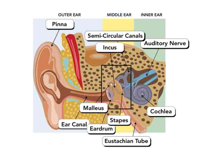
The outer ear, sometimes known as the external ear or auris externa, is the outermost portion of the ear’s anatomy. This anatomical structure is comprised of the pinna, the ear canal and the ear drum. Its primary purpose is to gather sound waves from the environment, concentrate them and direct them towards the more internal portions of the ear. 

The pinna (also known as the auricle or auricula) is the fleshy portion on the ear outside of the skull. The pinna’s main function is to collect sound waves from the external environment. It acts as a funnel for these waves, amplifying and directing them towards the ear canal. 

The ear canal (also known as the external auditory canal, external auditory meatus or the external acoustic meatus) is a tube-like structure that runs from the external pinna to the internal ear drum. Its primary function is to conduct sound waves in the air towards the ear drum, but it also produces cerumen (i.e., ear wax). Cerumen is a yellowish, waxy substance that serves to clean, lubricate and protect the ear canal.  

The ear drum (also known as the tympanic membrane) is a thin, cone shaped membrane that separates the outer ear from the middle ear. It stretches across the ear canal, and it begins the process of converting sound waves in the air into a form that the brain can comprehend.

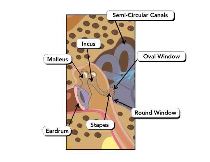
The middle ear is the portion of the ear between the ear drum and the oval window. It contains three bones, collectively the ossicles, which transfer vibrations from the ear drum into the inner ear in the form of compression waves. These bones are known commonly as the hammer, anvil and stirrup. The hollow space around the ossicles is the tympanic cavity. The whole structure is surrounded and protected by the tympanic portion of the temporal bone of the skull. 

The malleus, or hammer bone, is the first of the three ossicles. It is attached to the inner surface of the ear drum and connects to the incus. 

The incus, or anvil bone, is the second of the three ossicles. It bridges the gap between the malleus and the stapes, and it transfers sound vibrations between the two bones. 

The stapes, or stirrup bone, is the final of the three ossicles. It connects the incus to the oval window, and it transfers sound vibration to the inner ear. It is also the smallest and lightest bone in the human body.

The Eustachian tube (also known as the auditory tube or pharyngotympanic tube) is a tube that links the middle ear to the upper part of the pharynx, the nasopharynx. It is responsible for equalizing pressure in the middle ear and allowing mucus to drain out of the middle ear.  

Want to have a free hearing check?

Book an appointment

What is the inner ear?

The inner ear, sometimes known as the internal ear or auris interna, is the innermost portion of the ear. It consists mainly of the oval window, the cochlea, the semicircular canals and a portion of the auditory nerve. The inner ear also contains a central component to one's sense of balance, the vestibular system.

The most common category of hearing loss, sensorineural hearing loss (SNHL), occurs when there is a problem or damage to part(s) of the inner ear, typically the cochlea or the auditory nerve. While SNHL is usually permanent, wearing hearing aids can help mitigate the effects.

The oval window (also called the fenestra ovalis) is a connective tissue membrane that connects the inner ear to the middle ear. It is in direct contact with the stapes. The compression waves from the stapes travel through the oval window and into the fluid-filled cochlea. 

The cochlea is the portion of the inner ear most closely associated with hearing. It is a spiral-shaped, fluid-filled bone cavity. It contains the organ of Corti and the hair cells, both of which are integral in transforming compression waves from the middle ear into electrochemical impulses the brain can comprehend. 

The semicircular canals (also referred to as semicircular ducts) are three fluid-filled tubes in the inner ear. They are an important part of the vestibular system. The movement of fluid through the canals sends nerve impulses to the brain related to balance and an individual’s orientation in space. 

The auditory nerve (also known as the cochlear nerve or the acoustic neuron) is one of the two sections of the vestibulocochlear nerve. It carries neural impulses from the cochlea to the brain for interpretation. 
Woman leaning against wall

Avoid hearing problems

All of these different parts of the ear work together in a coordinated manner to deliver you the sense of sound. A problem with any one of the parts of the ear can have consequences on the whole process of hearing. If you or a loved one experience difficulty with your sense of hearing, make an appointment to visit a Miracle-Ear specialist today.

Get support and advice

Book an appointment online

Book now

Take a free online hearing test

Start test

Find a hearing aid center near you

Search now