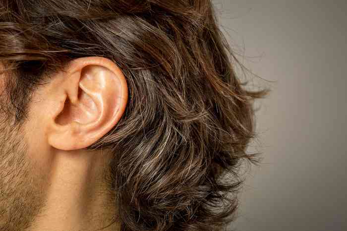
At first glance, the outer ear may seem pretty simple: it’s just a small curve of skin and cartilage on the side of your head. But what you see on the outside is only the beginning. This article will explore the anatomy, functions and common conditions afflicting the outer ear.
Our ears consist of external, middle and inner structures. The two terms for the outer ear are external ear or auris externa. This is the visible part of our hearing apparatus and the outermost portion of your ear’s anatomy.
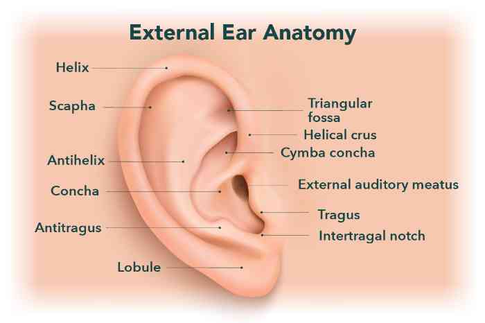
The anatomy of the ear is complex. There are three main parts of the outer ear: the pinna, the ear canal and the eardrum.1 Let’s dive into the specific function and definition of each of these parts of the outer ear.
The medical term for the outer ear is the pinna or auricle, which comes from the Latin word for “wing.” The pinna is the fleshy portion of the ear outside of the skull, made up of cartilage and skin. The pinna consists of several parts, including:
Antihelix, Antitragus, Concha, Helical crus, Helix, Lobe/lobule, Scapha, Tragus, Triangular fossa
The ear canal, or the external auditory canal, is a tube-like structure that runs from the external pinna to the internal eardrum. Its primary function is to conduct sound waves in the air toward the eardrum,2 and it is also responsible for producing earwax.
The tympanic membrane, also known as the eardrum, is what separates the outer and middle ear. It’s a thin layer of tissue with skin on the outside and lining on the inside.3 When sound waves hit the tympanic membrane, it vibrates. Those vibrations are sent to the inner ear, which are then transformed to electrical signals and then sent to your brain and interpreted as sound.
The outer ear gets its sense of feeling from several nerves working together, including the great auricular nerve, the lesser occipital nerve, the auriculotemporal nerve, the facial nerve, the vagus nerve and the glossopharyngeal nerve.
The purpose of the outer ear is to gather sound waves and funnel them into the inner ear, helping you hear more effectively.10
There are several important functions of the outer ear, including:
There are several common conditions and disorders that can affect the outer ear.
The outer ear can experience a few different problems, most of which are treatable.
Common conditions include swimmer’s ear (infection of the ear canal), earwax buildup and, more rarely, infections of the ear’s cartilage (perichondritis) or unusual growths on the outer ear (external audio exostosis, or surfer’s ear) or in the ear canal. These issues can cause pain, swelling or changes in hearing.
If you experience any of these symptoms, it might be a good time to check in with a healthcare provider.
Pain on the outer ear can come from several sources, such as:
When the outer ear is damaged or misshapen, surgery can sometimes restore its appearance or function. This may involve reshaping the ear, repairing injuries or closing a hole.
If you’re experiencing persistent pain or infection in your ears, whether the outer ear or inner structures, or if you’re noticing a decreased ability to hear, it’s time to see your healthcare professional.
For infections, visit your medical doctor. For any hearing-related concerns, you can book an appointment at your nearest Miracle-Ear location where you’ll receive a free hearing test from a licensed hearing care professional.
1 “Anatomy of the Ear.” National Human Genome Research Institute, elementsofmorphology.nih.gov/anatomy-ear.shtml. Accessed 11 Sept. 2025.
2 Szymanski, Alice, and Zachary Geiger. “Anatomy, Head and Neck, Ear.” U.S. National Library of Medicine, U.S. National Library of Medicine, 24 July 2023, www.ncbi.nlm.nih.gov/books/NBK470359/.
3 “Tympanic Membrane.” MedlinePlus, U.S. National Library of Medicine, 2 May 2024, medlineplus.gov/ency/imagepages/8993.htm.
4 Ginsberg, Lawrence E, and Susan A Eicher. “Great Auricular Nerve: Anatomy and Imaging in a Case of Perineural Tumor Spread.” AJNR. American Journal of Neuroradiology, U.S. National Library of Medicine, 21 Mar. 2000, pmc.ncbi.nlm.nih.gov/articles/PMC8174985/.
5 Yu, Megan, and Shu-Min Wang. “Anatomy, Head and Neck, Occipital Nerves.” StatPearls [Internet]., U.S. National Library of Medicine, 31 Oct. 2022, www.ncbi.nlm.nih.gov/books/NBK542213/.
6 Greenberg, Jacob S, and Michael J Breiner. “Anatomy, Head and Neck: Auriculotemporal Nerve.” StatPearls [Internet]., U.S. National Library of Medicine, 8 Aug. 2023, www.ncbi.nlm.nih.gov/books/NBK544240/.
7 “Role of the Vagus Nerve in Epilepsy: MedlinePlus Medical Encyclopedia Image.” MedlinePlus, U.S. National Library of Medicine, 16 Apr. 2025, medlineplus.gov/ency/imagepages/19252.htm.
8 Kaniusas, Eugenijus, et al. “Current Directions in the Auricular Vagus Nerve Stimulation I – A Physiological Perspective.” Frontiers in Neuroscience, U.S. National Library of Medicine, 9 Aug. 2019, pmc.ncbi.nlm.nih.gov/articles/PMC6697069/.
9 Thomas, Kathryn, et al. “Neuroanatomy, Cranial Nerve 9 (Glossopharyngeal).” StatPearls [Internet]., U.S. National Library of Medicine, 7 Nov. 2022, www.ncbi.nlm.nih.gov/books/NBK539877/.
10 Purves, Dale. “The External Ear.” Neuroscience. 2nd Edition., U.S. National Library of Medicine, 1 Jan. 1970, www.ncbi.nlm.nih.gov/books/NBK10908/.
11 “Overview: Outer Ear Infection.” InformedHealth.Org [Internet]., U.S. National Library of Medicine, 13 June 2023, www.ncbi.nlm.nih.gov/books/NBK279353/.
12 “Overview: Outer Ear Infection.” InformedHealth.Org [Internet]., U.S. National Library of Medicine, 13 June 2023, www.ncbi.nlm.nih.gov/books/NBK279353/.
13 “Outer Ear Infection: Research Summaries – What Helps If Earwax Builds Up?” InformedHealth.Org [Internet]., U.S. National Library of Medicine, 13 June 2023, www.ncbi.nlm.nih.gov/books/NBK279354/.

 Prevention is the key

 Find your closest Miracle-Ear center